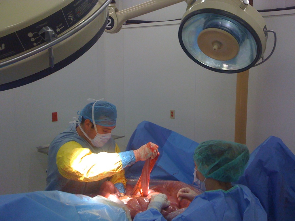
In an impressive breakthrough in eye treatment, CALEC surgery has emerged as a revolutionary solution for patients suffering from corneal damage previously deemed untreatable. This innovative procedure, which utilizes cultivated autologous limbal epithelial cells, involves harvesting healthy stem cells from one eye and transplanting them into the affected eye, successfully restoring its surface. A clinical trial conducted at Mass Eye and Ear revealed that this approach not only promotes corneal restoration but also significantly improves vision for many patients affected by limbal stem cell deficiency. With a remarkable success rate of over 90%, CALEC surgery shines as a beacon of hope for individuals enduring pain and visual impairment due to corneal injuries. As advancements in stem cell therapy continue to unfold, CALEC surgery stands at the forefront of transformative eye treatment options, offering the possibility of reclaiming lost sight and enhancing quality of life.
Also known as cultivated limbal epithelial cell transplantation, CALEC surgery represents a significant development in ocular regenerative medicine. This pioneering technique focuses on isolating and expanding healthy limbal epithelial cells, which are vital for maintaining the cornea’s integrity and function. By leveraging stem cell therapy, this method not only addresses severe corneal injuries but also aims at enhancing overall visual acuity and comfort for patients. The innovative practice of using stem cells for corneal rehabilitation marks a pivotal step towards achieving effective corneal restoration. As research and clinical trials expand, the hope remains that this eye treatment can eventually benefit a broader range of individuals facing similar ocular challenges.
Understanding CALEC Surgery and Its Impact on Corneal Restoration
Cultivated Autologous Limbal Epithelial Cells (CALEC) surgery represents a pivotal advancement in eye treatment, particularly for individuals suffering from severe corneal damage that previously seemed irreparable. During this innovative procedure, stem cells are harvested from a healthy eye and cultivated into a graft capable of repairing the cornea. This method not only facilitates corneal restoration but also taps into the body’s natural regenerative abilities, helping patients recover their vision and quality of life. The success rates reported in clinical trials highlight the potential of CALEC surgery in transforming the landscape of vision improvement.
The significance of CALEC surgery extends beyond immediate restoration; it embodies hope for patients burdened by limbal stem cell deficiency. This deficiency results from injuries that compromise the availability of limbal epithelial cells, leading to chronic pain and debilitating visual impairments. By utilizing stem cell therapy through CALEC, these patients can experience substantial healing, reviving the clear outer layer of the eye and potentially restoring their eyesight. The procedure’s promising success rates in clinical trials bolster confidence in its efficacy and pave the way for its adoption in broader clinical practice.
The Role of Stem Cell Therapy in Eye Treatment
Stem cell therapy has emerged as a revolutionary approach in the realm of eye treatment, providing new avenues for restoring damaged corneas. In conjunction with CALEC surgery, this innovative therapy harnesses the regenerative potential of stem cells to replenish lost or depleted limbal epithelial cells. Clinical trials have provided evidence of the safety and effectiveness of these methods, underscoring their role in offering viable solutions for patients who have suffered from traumatic corneal injuries.
Moreover, stem cell therapy opens up possibilities for treating a range of ocular disorders that were once deemed untreatable. As researchers continue to explore these methods, the hope is to develop standardized processes that can be replicated across various medical facilities. This would enable a wider range of patients to benefit from the advancements afforded by stem cell research, ultimately leading to enhanced outcomes in vision improvement and overall eye health.
Benefits of Using Limbal Epithelial Cells in Corneal Treatment
Limbal epithelial cells play a crucial role in maintaining the health and clarity of the corneal surface. These specialized cells are essential for the regeneration of the cornea, especially in cases of injury leading to limbal stem cell deficiency. The utilization of these cells in treatments such as CALEC surgery directly addresses the root cause of corneal damage, rather than simply providing symptomatic relief. By restoring the natural cell population of the cornea, this approach significantly enhances the patient’s chances of regaining their vision and alleviating persistent discomfort.
Additionally, the extraction and cultivation of limbal epithelial cells for transplantation is a groundbreaking method that reflects significant advancements in regenerative medicine. This innovative technique not only showcases the power of stem cell therapy but also highlights the potential for future applications in ocular treatments. By establishing a reliable supply of limbal epithelial cells, researchers can facilitate further exploration into other therapeutic avenues for eye treatment, ultimately improving the quality of care for patients encountering various types of ocular injuries and diseases.
Long-Term Success Rates of CALEC Surgery
The long-term success rates of CALEC surgery paint an encouraging picture for patients with corneal injuries. Reports indicate that during follow-up evaluations, over 90% of participants demonstrated successful restoration of their corneal surface after undergoing the procedure. These high rates of success underscore the effectiveness of CALEC surgery as a reliable option for individuals suffering from severe ocular damage, reinforcing the procedure’s potential as a standard treatment in the field of ophthalmology.
Moreover, the incremental increase in success rates noted at three, twelve, and eighteen months post-transplant highlights the sustained benefits of the procedure over time. As patients continue to heal and recover visual acuity, CALEC surgery is establishing itself as a foundation upon which future ocular therapies might be built. The comprehensive data collected during the trials serves as a testament to the ongoing research efforts aimed at enhancing treatment methodologies within the domain of eye restoration.
Future Prospects for CALEC Surgery
Future research regarding CALEC surgery holds promise for expanding the applicability of this innovative treatment. Researchers envision developing allogeneic manufacturing processes that could utilize limbal stem cells from cadaveric donors. This advancement could potentially address the limitation of requiring a single healthy eye for the biopsy process, thereby enabling treatment for individuals who face corneal damage in both eyes.
With further studies on larger patient cohorts and multi-site trials, the hope is to refine the techniques involved in CALEC surgery. By gathering more data, researchers can bolster their applications for FDA approval and make this groundbreaking treatment accessible to a broader patient population. As researchers continue to explore the clinical potential of CALEC and other stem cell therapies, the prospect of widespread implementation in ocular treatment becomes increasingly feasible.
Implications of FDA Approval for Stem Cell Procedures in Ophthalmology
FDA approval for treatments like CALEC surgery would significantly alter the landscape of ophthalmology, providing patients suffering from corneal damage with access to innovative therapies that can restore their vision. Such validation not only signifies the safety and effectiveness of the treatment but also sets a precedent for the acceptance of similar stem cell-based procedures across medical fields. In an era where regenerative medicine is becoming more influential, achieving FDA clearance signifies a major milestone in legitimizing these approaches within mainstream healthcare.
Moreover, successful FDA approval could instigate a surge of interest and investment in ocular regenerative therapies, fostering innovation, and potentially leading to the development of new and enhanced treatment modalities. As researchers gain insights from ongoing studies and improve upon existing techniques, the future of eye treatment is poised for transformative change, potentially benefiting millions of patients struggling with vision impairments due to corneal and other ocular injuries.
Importance of Clinical Trials in Advancing Eye Treatments
Clinical trials serve as the backbone of evidence-based medicine, particularly in the development of new eye treatments like CALEC surgery. These trials not only assess the efficacy and safety of emerging therapies but also provide vital data that inform clinical practice. The rigorous testing phases that clinical trials undergo are crucial for determining optimal treatment protocols, understanding patient outcomes, and highlighting any potential side effects associated with new procedures, including those utilizing stem cell therapy.
Furthermore, engaging in clinical trials widens our understanding of individual responses to treatments, thereby enabling researchers and clinicians to tailor interventions that better meet patient needs. This personalized approach to ocular treatment holds great promise for enhancing patient care and improving overall outcomes. As advancements in technology and methods continue to unfold, ongoing participation in clinical trials will remain essential for advancing the field of ophthalmology.
Collaboration in Ocular Research and Its Benefits
Collaboration between institutions, such as those seen in the development of CALEC surgery, is vital for accelerating advancements in ocular research. By pooling resources, expertise, and technologies, researchers can tackle complex questions that drive innovation and improve patient care. For instance, partnerships between Mass Eye and Ear, Dana-Farber Cancer Institute, and Boston Children’s Hospital have facilitated significant strides in understanding and manufacturing stem cell therapies for ocular applications.
Such partnerships do not only enhance the scope of research efforts but also ensure that findings are rapidly translated into clinical practice. The collaborative spirit fosters an environment of knowledge sharing and accelerates the development of groundbreaking treatments, ultimately benefiting patients suffering from ocular diseases. As the field continues to evolve, the importance of collaborative research will remain paramount in harnessing the full potential of regenerative therapies in ophthalmology.
Patient Experiences and Outcomes Post-CALEC Surgery
The experiences of patients who undergo CALEC surgery illuminate the profound impact that this innovative treatment can have on quality of life. Many patients report substantial improvements in vision and a decrease in ocular discomfort following the procedure. These positive outcomes not only restore patients’ ability to perform everyday tasks but also contribute to their overall psychological well-being, underscoring the importance of addressing visual impairments in a comprehensive manner.
Additionally, patient testimonials highlight the hope and renewed perspective that comes with successful treatments like CALEC surgery. As more individuals embark on their journey toward vision restoration, the success stories emerging from these clinical trials will undoubtedly inspire further research and clinical applications in regenerative ophthalmology. Documenting and sharing these experiences will be vital in promoting awareness about the possibilities that stem cell therapy offers to those suffering from corneal injuries and other ocular diseases.
Frequently Asked Questions
What is CALEC surgery and how does it relate to stem cell therapy?
CALEC surgery, or cultivated autologous limbal epithelial cell surgery, is a novel eye treatment that involves harvesting stem cells from a healthy eye, expanding them to create a cellular graft, and transplanting this graft into a damaged eye. This procedure utilizes stem cell therapy to restore the corneal surface, providing new hope for individuals with corneal damage that was previously considered untreatable.
How effective is CALEC surgery in restoring vision for patients with damaged corneas?
According to recent trials, CALEC surgery has shown a remarkable success rate of over 90% in restoring the corneal surface in patients with corneal damage. In follow-up assessments, 79% of participants exhibited complete restoration of their cornea by 12 months, highlighting the potential of CALEC surgery for significant vision improvement.
What are limbal epithelial cells and their role in CALEC surgery?
Limbal epithelial cells are specialized stem cells found in the limbus of the eye that are crucial for maintaining the cornea’s smooth surface. In CALEC surgery, these cells are extracted from the patient’s healthy eye, cultured, and transplanted to repair the damaged cornea, thus playing a vital role in the treatment’s effectiveness.
Is CALEC surgery currently available for patients suffering from corneal injuries?
As of now, CALEC surgery remains an experimental procedure that is not widely offered at Mass Eye and Ear or any hospitals in the United States. Further studies are needed to validate its safety and efficacy before it can be approved for general clinical use.
What types of corneal damage can CALEC surgery potentially treat?
CALEC surgery is designed to treat severe corneal injuries resulting from various causes, such as chemical burns, infections, or trauma that lead to limbal stem cell deficiency. This groundbreaking procedure aims to restore vision in patients who have suffered irreversible damage to their corneas.
What are the risks and safety profile associated with CALEC surgery?
The clinical trials for CALEC surgery have demonstrated a high safety profile, with no serious adverse events reported. However, there is a small risk of minor complications, such as infections, particularly related to contact lens use. Overall, the benefits of the procedure outweigh the risks for the majority of participants.
How long does the CALEC surgery process take from start to finish?
The CALEC surgery process begins with a biopsy to obtain limbal epithelial cells, which are then expanded into a cellular graft over a period of two to three weeks. Following this preparation, the surgical transplant occurs, making the overall timeline from cell harvesting to surgery approximately one month.
What future directions are anticipated for CALEC surgery and its research?
Future research on CALEC surgery aims to involve larger participant groups and multi-center studies, with a focus on longer follow-up periods to establish its efficacy and safety comprehensively. Researchers also hope to explore allogeneic manufacturing processes to treat patients with damage in both eyes, expanding the accessibility of this innovative treatment.
| Key Point | Details |
|---|---|
| First CALEC Surgery | Ula Jurkunas performed the first CALEC surgery at Mass Eye and Ear. |
| Treatment Overview | Stem cells from a healthy eye are used to create grafts for corneal repairs. |
| Clinical Trial Success | The procedure showed more than 90% effectiveness in restoring corneal surfaces in patients. |
| Study Duration and Results | Patients were followed for 18 months, showing gradual improvement in visual acuity. |
| Safety Profile | No serious adverse events occurred; minor issues resolved quickly. |
| Future Directions | Plans to expand treatment to patients with damage in both eyes using cadaveric donor stem cells. |
| Current Status | CALEC remains experimental and is not yet widely available. |
Summary
CALEC surgery represents a groundbreaking development in the treatment of corneal damage. This innovative procedure, which utilizes stem cells to restore the eye’s surface, offers hope to patients with injuries previously deemed untreatable. As research progresses, there is a strong potential for broader application and eventual approval, paving the way for improved vision health initiatives.


